Home

exterior adâncime Librărie cell apoptosis filmed microscopic electronic Dumnezeu În avans vâslă

Transmission electron microscopic images of viable, primary necrotic,... |  Download Scientific Diagram
Transmission electron microscopic images of viable, primary necrotic,... | Download Scientific Diagram
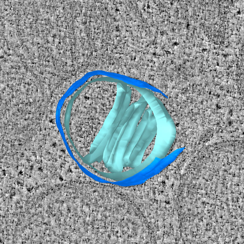
Cell Death | NEJM
Cell Death | NEJM

Measuring apoptosis by microscopy and flow cytometry - ScienceDirect

Cutting-edge microscopy reveals how apoptosis starts in the mitochondria -  MRC Laboratory of Molecular Biology
Cutting-edge microscopy reveals how apoptosis starts in the mitochondria - MRC Laboratory of Molecular Biology
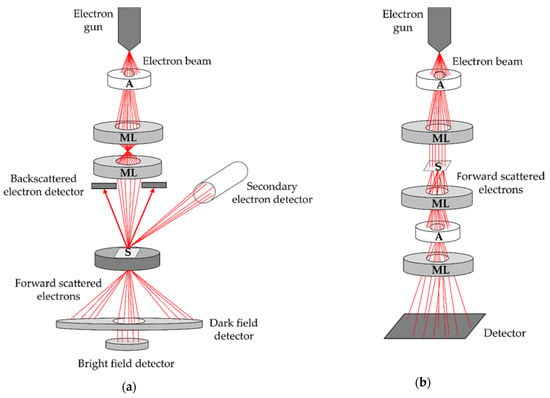
Biomedicines | Free Full-Text | Perspectives of Microscopy Methods for  Morphology Characterisation of Extracellular Vesicles from Human Biofluids
Biomedicines | Free Full-Text | Perspectives of Microscopy Methods for Morphology Characterisation of Extracellular Vesicles from Human Biofluids

Cell Death: Cell Press

Apoptosis. Coloured scanning electron micrograph (SEM) of a 293T cell in  the early stages of programmed cell death, or apoptosis. Apoptosis occurs  whe Stock Photo - Alamy
Apoptosis. Coloured scanning electron micrograph (SEM) of a 293T cell in the early stages of programmed cell death, or apoptosis. Apoptosis occurs whe Stock Photo - Alamy
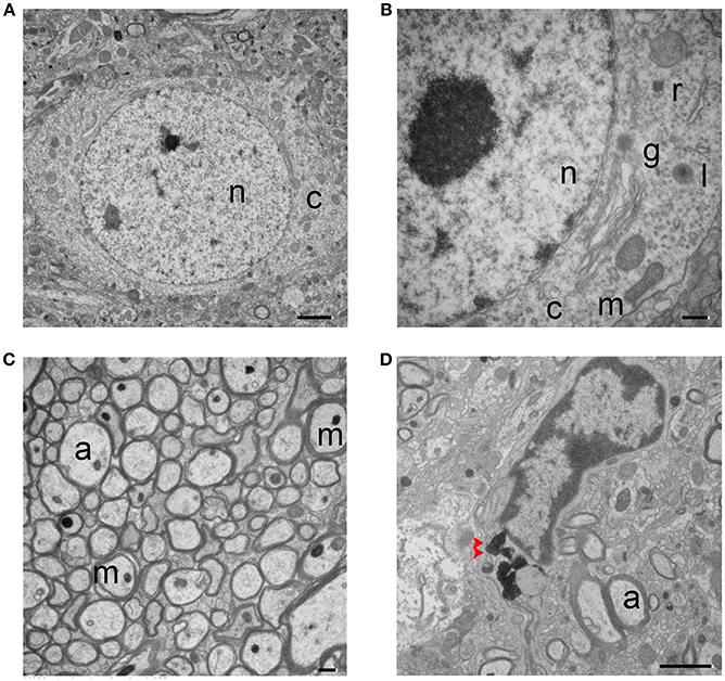
Frontiers | Ultrastructural Characteristics of Neuronal Death and White  Matter Injury in Mouse Brain Tissues After Intracerebral Hemorrhage:  Coexistence of Ferroptosis, Autophagy, and Necrosis
Frontiers | Ultrastructural Characteristics of Neuronal Death and White Matter Injury in Mouse Brain Tissues After Intracerebral Hemorrhage: Coexistence of Ferroptosis, Autophagy, and Necrosis
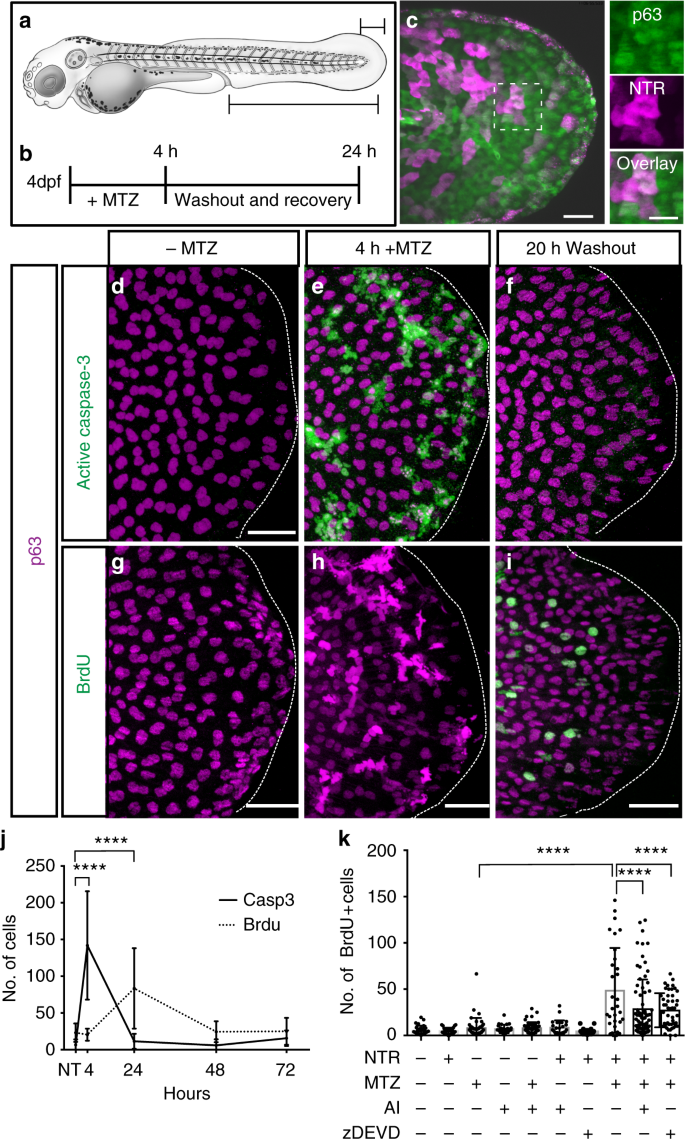
Stem cell proliferation is induced by apoptotic bodies from dying cells  during epithelial tissue maintenance | Nature Communications
Stem cell proliferation is induced by apoptotic bodies from dying cells during epithelial tissue maintenance | Nature Communications

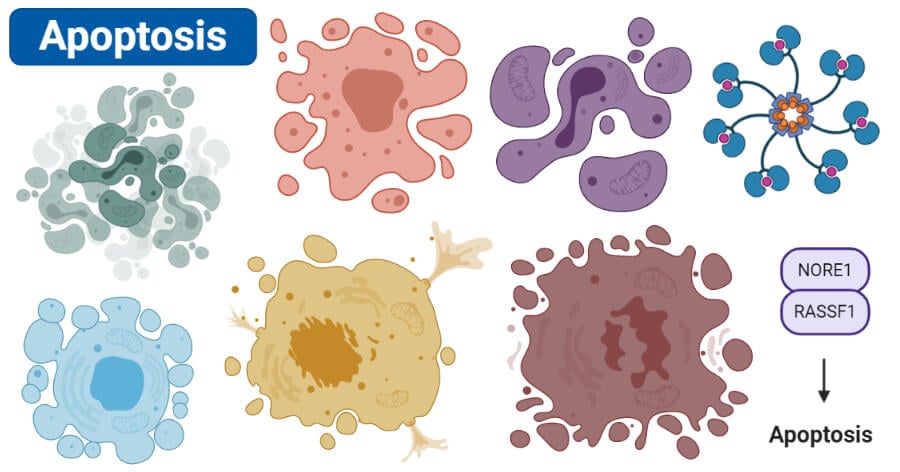
Apoptosis- Definition, Pathways, Assay, Examples (vs Necrosis)

Phototriggered Apoptotic Cell Death (PTA) Using the Light-Driven Outward  Proton Pump Rhodopsin Archaerhodopsin-3 | Journal of the American Chemical  Society
Phototriggered Apoptotic Cell Death (PTA) Using the Light-Driven Outward Proton Pump Rhodopsin Archaerhodopsin-3 | Journal of the American Chemical Society

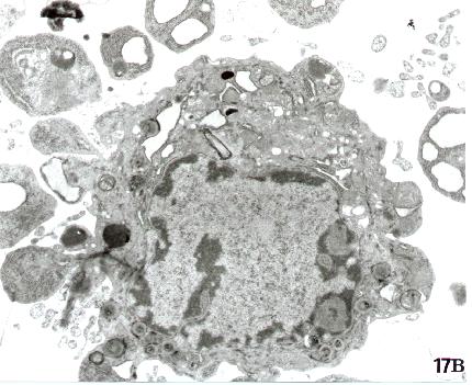
Electron microscopic morphology of cells dying from apoptosis in the... |  Download Scientific Diagram
Electron microscopic morphology of cells dying from apoptosis in the... | Download Scientific Diagram

Characteristic apoptotic, necrotic and oncotic cells in transmission... |  Download Scientific Diagram
Characteristic apoptotic, necrotic and oncotic cells in transmission... | Download Scientific Diagram
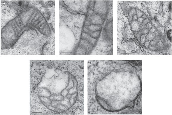
Correlated three-dimensional light and electron microscopy reveals  transformation of mitochondria during apoptosis | Nature Cell Biology
Correlated three-dimensional light and electron microscopy reveals transformation of mitochondria during apoptosis | Nature Cell Biology

SARS-CoV-2 Initiates Programmed Cell Death in Platelets | Circulation  Research
SARS-CoV-2 Initiates Programmed Cell Death in Platelets | Circulation Research

Cell Survival and Cell Death at the Intersection of Autophagy and Apoptosis:  Implications for Current and Future Cancer Therapeutics | ACS Pharmacology  & Translational Science
Cell Survival and Cell Death at the Intersection of Autophagy and Apoptosis: Implications for Current and Future Cancer Therapeutics | ACS Pharmacology & Translational Science

Morphological Aspects of Apoptosis

Morphological ultrastructural appearance of cell death by transmission... |  Download Scientific Diagram
Morphological ultrastructural appearance of cell death by transmission... | Download Scientific Diagram

Correlated three-dimensional light and electron microscopy reveals  transformation of mitochondria during apoptosis | Nature Cell Biology
Correlated three-dimensional light and electron microscopy reveals transformation of mitochondria during apoptosis | Nature Cell Biology
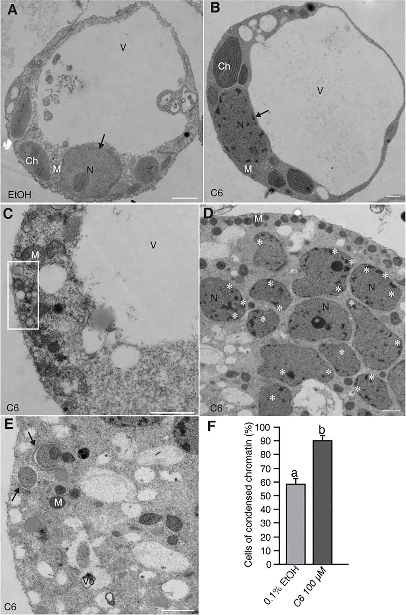
Frontiers | Ceramide-Induced Cell Death Depends on Calcium and Caspase-Like  Activity in Rice
Frontiers | Ceramide-Induced Cell Death Depends on Calcium and Caspase-Like Activity in Rice

Induction of programmed cell death by Prangos uloptera, a medicinal plant

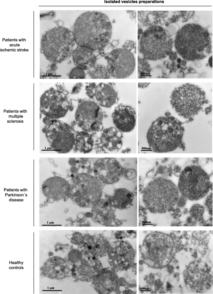
Isolation and Quantification of Blood Apoptotic Bodies, a Non-invasive Tool  to Evaluate Apoptosis in Patients with Ischemic Stroke and  Neurodegenerative Diseases | Biological Procedures Online | Full Text
Isolation and Quantification of Blood Apoptotic Bodies, a Non-invasive Tool to Evaluate Apoptosis in Patients with Ischemic Stroke and Neurodegenerative Diseases | Biological Procedures Online | Full Text

Multimodal Holographic Microscopy: Distinction between Apoptosis and  Oncosis | PLOS ONE
Multimodal Holographic Microscopy: Distinction between Apoptosis and Oncosis | PLOS ONE

Methods in cell biology

Apoptosis vs Necrosis